CERSI Conference Spotlights Need for Mass Spectrometry in Drug Development
Researchers from academia, government, and industry examine current challenges surrounding mass spectrometry as a drug development tool, including its status of adoption by industry and potential regulatory implications.

By Malissa Carroll
April 16, 2014
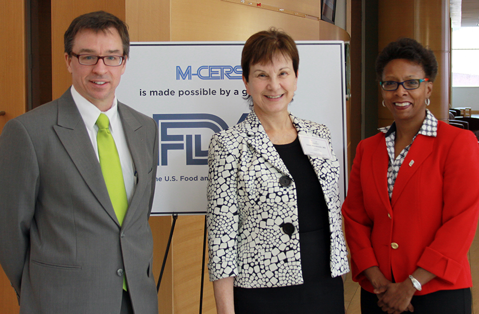
On April 8, the University of Maryland School of Pharmacy welcomed researchers from academia, government, and industry to “MALDI-Mass Spectrometry Imaging of Drug Metabolism,” a conference sponsored by the University of Maryland Center of Excellence in Regulatory Science and Innovation (M-CERSI) and the Food and Drug Administration (FDA). Organized by Maureen Kane, PhD, assistant professor in the Department of Pharmaceutical Sciences (PSC) at the School, this one-day conference explored the benefits and challenges associated with using matrix-assisted laser desorption ionization-mass spectrometry imaging (MALDI-MSI) as a tool in the drug development process.
“As a department, PSC encompasses all fields within drug discovery and development,” said Andrew Coop, PhD, professor and chair of PSC, who welcomed conference attendees to the School. “Mass spectrometry plays an integral role in almost all aspects of research conducted within our department. We currently maintain 15 mass spectrometers in our state-of-the-art Mass Spectrometry Center, which allows our researchers to continue pursuing the School’s vision of advancing scientific knowledge across the spectrum of drug discovery, health services, and practice-based and translational research in the state of Maryland and beyond.”
MALDI-MSI is an analytical technique that allows researchers to spatially characterize the molecular content of intact tissues or cell cultures using mass spectrometry. To perform MALDI-MSI, researchers fire a laser at a tissue sample that has been placed on a target and loaded with a special matrix. The laser disrupts the tissue, releasing ions that are captured and analyzed by the mass spectrometer. These ions show researchers which drugs, metabolites, and other molecules are present in the tissue.
MALDI-MSI has been used to follow drug metabolism and map drug distribution on a cellular level, as well as evaluate cellular changes or side effects associated with a particular drug. At the conference, speakers Stephen Castellino, PhD, US director of drug metabolism and pharmacokinetics at GlaxoSmithKline (GSK); Brendan Prideaux, PhD, visiting researcher at the Public Health Research Institute at Rutgers New Jersey Medical School; and Kane presented how they are currently using this effective technique in their research.
Delivering the conference’s kick-off presentation, Castellino spoke about using MALDI-MSI in his work with other GSK researchers to develop safe and effective dermal drugs. One drug on which he collaborated was designed to treat hyperhidrosis – a condition in which individuals experience profuse sweating or perspiration.
“All dermal medications must be formulated in such a way that they are able to move from the epidermis to the dermis, but no further, as that would increase patients’ risk for systemic exposure,” said Castellino. “At GSK, MALDI-MSI has made a dramatic impact in both drug discovery and development. It has changed our views on tissue distribution, giving us the opportunity to analyze those distributions over a pre-determined period of time. It’s also an integrated modality and involves scientists from a wide range of fields working together to pool their data and better understand drug pathways and mechanisms.”
Prideaux presented his research using MALDI-MSI to optimize drug discovery and development for tuberculosis, an often severe bacterial infection that currently affects one-third of the world’s population. Although a number of drug regimens exist to treat tuberculosis, successful treatment typically requires patients to spend six to 24 months in the hospital.
“In an active tuberculosis infection, patients develop granulomas in their lung tissue,” said Prideaux. “We used MALDI-MSI to see whether the drugs used to treat this illness are able to penetrate these granulomas and reach the different areas on which they need to act to be effective. Our imaging illustrated that one drug was able to penetrate very well into the lesion. This truly shows the value of MALDI-MSI. It gives us detailed spatial information to which we otherwise would not have access using standard quantification methods.”
Kane later spoke about her participation in the Medical Countermeasures Against Radiological and Nuclear Threats (MCART) Consortium – a large, federally-funded, multi-institutional program established to develop medical countermeasures against the lethal exposure to ionizing radiation. As co-director of the School’s Mass Spectrometry Center, Kane uses her expertise in MALDI-MSI to help the consortium identify and validate new biomarkers of radiation injury, recovery, and intervention in the hematopoietic, gastrointestional, and lung systems.
“Ionized radiation can cause a wide range of injuries, which can be acute or delayed,” said Kane. “Because of the complexity of this injury, we are using MALDI-MSI to help develop new therapies that will target key biological processes and mechanisms within the tissue. The spatial information provided by MALDI-MSI has proven to be incredibly important in our work. We have also found this technique to be very accessible, allowing us to apply our analytical skills to address a wide range of important issues associated with this type of injury.”
Moving beyond the discussion of applications for MALDI-MSI, Per Andren, PhD, senior lecturer in the Department of Pharmaceutical Biosciences at the University of Uppsala in Sweden, engaged the audience in a discussion about the unique advantages associated with this innovative imaging technique. He spoke about a collaboration in which he participated with a major drug company to identify crystals in kidney tissues using MALDI-MSI. “Using mass spectrometry, we were able to detect and identify even the smallest crystals in the tissue. The histologists who examined the tissue before us had not seen those particular crystals,” he noted.
Additional speakers at the conference included Lisa Cazares, PhD, proteomic scientists for the Army Research Institute of Infectious Disease, who spoke about the different tissue fixation methods that researchers can employ to prepare tissue samples for MALDI-MSI, and Michelle Reyzer, PhD, research assistant professor of biochemistry at Vanderbilt University, who spoke about additional applications for MALDI-MSI in the analysis of small molecules.
Following the presentations, Kane led the audience in a panel discussion that address how MALDI-MSI could be used to bridge biology and chemistry in drug development, how unique cell-based models could be used to study toxicity, the importance of using diverse data sources to aid in the prediction of drug action, and the accessibility of MALDI-MSI as an imaging technique and its use to study drug distribution. “The presentations delivered today were excellent. Hopefully, this discussion will help guide future regulations surrounding the use of MALDI-MSI in drug discovery and development at the FDA,” she said.
Related News Stories
CERSI Symposium Highlights Use of Biomarkers in Drug Development
August 28, 2015
More than 500 researchers from academia, government, and industry gather to gain perspective on biomarker development and the application of biomarkers in preclinical and clinical research.
CERSI Conference Spotlights New Applications for Top-Down Mass Spectrometry
June 30, 2014
Researchers from academia, government, and industry examine benefits and new methods associated with emerging technology.

SOP Joins International Group to Advance Treatments for Radiation Injuries
January 31, 2013
The Mass Spectrometry facility at the University of Maryland School of Pharmacy will lead investigation to identify and characterize new biomarkers of radiation damage.